
-
- BoldMast EnanMast PropNandro DecaNandro PPPrimobolanSustanonTest CypTest EnanTest propTren AceateTren BlendTren ETren Hex
- AnadrolAnavarArimidexClenbuterolClomidDianabolProvironStanozololT3Tadalifil
Mechanical stress, skeletal muscle growth, and intrinsic adaptation mechanisms
Views: 2 Author: Site Editor Publish Time: 2024-07-10 Origin: Site
Mechanical stress is one of the main inducing factors for muscle hypertrophy (the other two being metabolic stress and muscle injury, as seen in previous articles), which is not really news. There is evidence to suggest that as early as the 7th century BC, humans have realized that mechanical stress can promote body growth. For humans, resistance training is widely known as the main means of inducing muscle growth: studies have shown that 8-16 weeks of resistance training can increase the volume/mass of skeletal muscles by 5-20%. In animal experiments, the use of collaborative ablation (which refers to the surgical removal of the gastrocnemius and soleus muscles of experimental animals, causing the remaining plantar muscles to be forced to overload) technology can promote the doubling of muscle mass in experimental mice in just two weeks [3]! Human and animal research has provided us with a deeper understanding of the effects and underlying mechanisms of mechanical stress on skeletal muscle hypertrophy.
Basic structure and background knowledge of skeletal muscles
At the macro level, skeletal muscles are connected to the bone through tendons and achieve their contractile function by providing power and angle changes to the muscles. From a microscopic perspective, skeletal muscle is enveloped by the connective tissue of the outer membrane, while below the outer membrane lies the muscle fiber bundle, which is enveloped by the connective tissue of the perimuscular membrane. (See the "Microscopic Perspective of Muscle Fibers" diagram)
In the theoretical model, we assume that each muscle fiber is parallel to the longitudinal axis of the muscle (i.e. the line connecting the starting and ending points of the muscle), but in real muscles, the muscle bundle (=muscle fiber) is not always parallel to the longitudinal axis of the muscle, but deviates from the longitudinal axis at a certain angle, which is called the feather angle
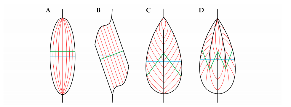
(There are four main ways to arrange feather shaped angles: A: spindle shape (the anatomical cross-sectional plane of muscle fibers overlaps with their physiological cross-sectional plane), B: single angle (there is an angle between the anatomical cross-sectional plane of muscle fibers and their physiological cross-sectional plane), C: double angle (there is an angle between the anatomical cross-sectional plane of muscle fibers and their physiological cross-sectional plane), D: multiple angles (there are two or more angles between the anatomical cross-sectional plane of muscle fibers and their physiological cross-sectional plane). The anatomical cross-section of muscle fibers is the plane perpendicular to the (macroscopic) longitudinal axis of the muscle; Physiological cross-section of muscle fibers=plane perpendicular to the longitudinal axis of muscle fibers (microscopic))
Take a single muscle fiber as an example to examine the internal structure of muscle fibers. Muscle fibers are a type of multinucleated cells, which are surrounded by the connective tissue of the endometrium on the outside. There are various stromal cells on the outside of muscle fibers, such as fibroblasts, immune cells, pericytes, and fibro adipogenic promoters.
Satellite cells, located between the endometrium and sarcolemma (a type of connective tissue that wraps around muscle fibers), are important cells responsible for muscle growth, repair, and more. The components enclosed by the muscle membrane are called sarcoplasm. According to research, about 80% of the space in the muscle is filled with parallel rod-shaped structures called myofibrils. The myofibrils are composed of a long string of components that can generate power (association: string Tomatoes on sticks), called sarcomere. The sarcomeres are surrounded by the sarcoplasmic reticulum, and there are mitochondria between adjacent sarcomeres (myofibrils).
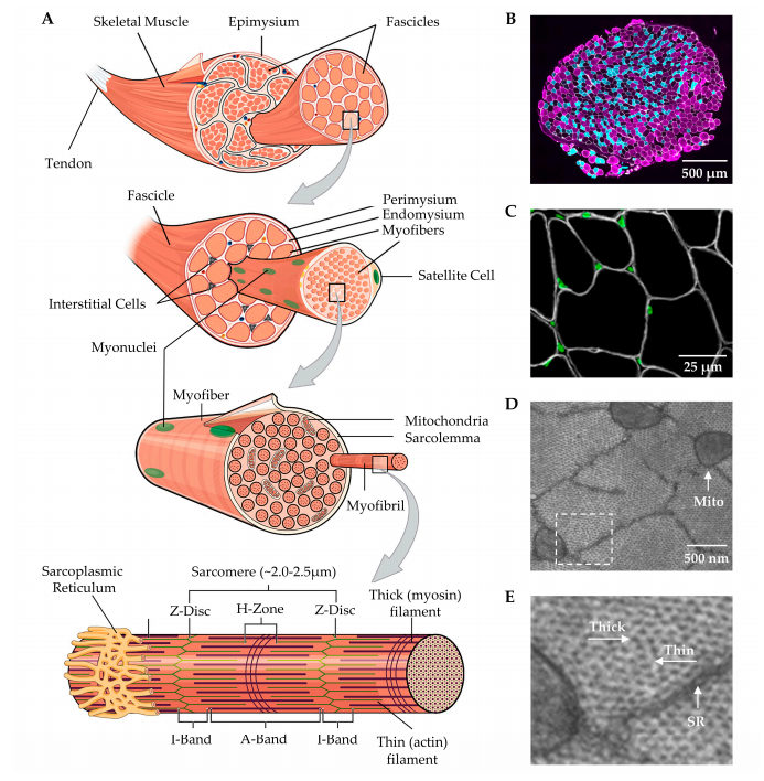
The sarcomere exerts its contraction function through the relative sliding between the coarse and fine filaments, and this process can be described by the theory of sarcomere sliding. As shown in the figure, the sarcomere is composed of a Z-disk, an I-band containing fine filaments (actin), and an A-band containing coarse filaments (myosin). The relative movement between the coarse and fine filaments in several muscle segments is manifested microscopically as the Z-disk approaching the H region (as shown in the figure above), while macroscopically it is manifested as muscle tissue shortening, i.e. muscle contraction.
Muscle fibers can be roughly divided into two types based on their contraction properties. One type is called slow contracting muscle fibers, which are characterized by severe dependence on oxidative metabolism, slow contraction speed, and anti fatigue properties; The other type relies on anaerobic glycolysis, which results in fast contraction and fatigue. According to the isomers of myosin heavy chain (MHC) in different muscle fibers, they can be classified into three main types: type I (slow oxidation), IIA (rapid oxidation), and IIB (rapid glycolysis).
reference material:
1、Todd, J. From Milo to Milo: A History of Barbells, Dumbells, and Indian Clubs. Iron Game History 1995, 3,4–16.
2、Abe, T.; Kojima, K.; Kearns, C.F.; Yohena, H.; Fukuda, J. Whole body muscle hypertrophy from resistance training: distribution and total mass. Br. J. Sports Med. 2003, 37, 543–545
3、Lowe, D.A.; Alway, S.E. Animal models for inducing muscle hypertrophy: are they relevant for clinical applications in humans? J. Orthop. Sports Phys. Ther. 2002, 32, 36–43
4、MacDougall, J.D.; Sale, D.G.; Elder, G.C.; Sutton, J.R. Muscle ultrastructural characteristics of elite powerlifters and bodybuilders. Eur. J. Appl. Physiol. Occup. Physiol. 1982, 48, 117–126
5、Fry, A.C. The role of resistance exercise intensity on muscle fibre adaptations. Sports Med. 2004, 34, 663–679


